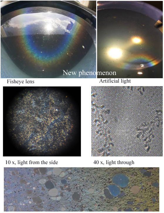
 Some time ago Jari Piikki posted me a couple of drops of the surface film that produced his colourful corona. I mixed it in a bucket full of tap water with also some forest litter added. After a week or so there appeared a bluish film on water surface. Closer inspection with point-like light revealed more colors. It was actually a large corona, with blue inside, and green and red on the outer edge.
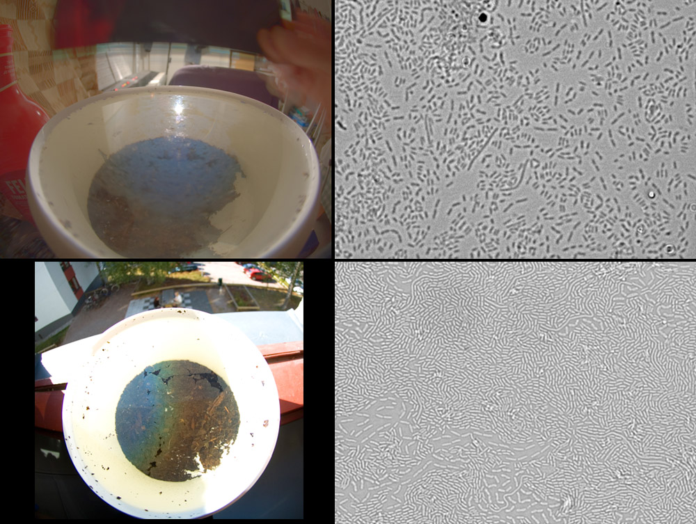
Only a segment of the corona could be seen in the small bucket. This is shown in the upper left image. Unlike the peculiar corona described by Piikki, this one has true blue aureole starting right from the reflection image of the lamp. Outside the blue some green and perhaps red maybe distinguished – visually these colors were clear. Upper right image is microphotograph of the film. It consisted of bacteria.
Day after taking the upper photographs the phenomenon in the bucket changed. Now there was similar corona as what Piikki photographed, only less bright. This is shown in lower left. There was no aureole, but instead a dark area surrounded the light source. This also was consistent with Piikki’s observation. Microphotograph of the film on this stage is shown on the lower right.
The most conspicuous difference in the bacterial level between the two stages is the crowding – there are more bacteria in the latter stage. Is this somehow related to the changes in the phenomena? Perhaps Minnaert can give an answer. In the chapter about coronas seen in windows, he describes how these coronas have an aureole replaced by dark area. He speculates the dark area forms because in the two dimensional plane of the window surface the droplets can be evenly spaced – a situation which can not happen when particles are freely floating in the air.
Now, if one looks at the lower right microphotograph, one can imagine that these bacteria indeed are spaced more evenly as compared to the upper photo. In the Piikki’s microphotograph, which looks like a maze, the bacterial film might consist solely on the long strands that are seen in small numbers also in my lower right photo. When packed next to each other, these kind of strands would possibly be very evenly spaced, thus perhaps contributing to the stronger corona. Piikki’s photograph is, however, too much out of focus to be sure of this interpretation.
The bacteria species could be Nevskia ramosa, which is well known neustonic (water surface inhabiting) bacteria. Genomic fingerprinting would be needed for confirmation. For the last note it should be said that the inverted colors between the two microphotographs are due to differences in focus. The upper image is a bit out of focus and thus the bacteria shows as dark spots.
No comments:
Post a Comment